Non-destructive survey of bookbinding structure using X-ray radiation
Keywords: X-ray radiation, radiography, book collection, bookbinding, visualisation
Ing. Petra Vávrová, Ph.D., Mgr. Jitka Neoralová, Dana Hřebecká, Ing. Kristýna Kohoutová, MgA. Anna Kulíčková, Bc. Marie Matysová, Daniela Popelková, Tomáš Blecha / Odbor ochrany knihovních fondů, Oddělení vývoje a výzkumných laboratoří, Národní knihovny České republiky (Division of preservation of library collections, Department of development and experimental laboratories of the National Library of the Czech Republic), Klementinum 190, 110 00 Prague 1, Czech Republic
Introduction
Bookbinding represents a rich source not only of textual and visual information, but it is also a physical document of art and craft creation of a binder, contemporary trend of technology of production, and last but not least a document of history of proper existence reverberating in defects from wear and tear and from natural decomposition of materials. Technology of making books and their damages are often hidden beneath layers of materials, and it is possible to find them out without invasive intervention only with difficulty. By the help of through illuminative technology making use of X-rays, it is possible to penetrate not destructively below the surface of outer layers to inner structures and structural elements of bookbinding. Within the frame of the grant project of the Ministry of Culture NAKI II with name "Utilization of imaging methods for study of hidden information in books", the workplace of the Department of development and experimental laboratories of the National library CR (hereinafter also NL CR) was equipped with an x-ray box, a part of which is an X-ray source and digital detector of flat panel type. Information on used technologies, materials, and their conditions are fundamental also for historic, artistic and scientific understanding of bookbinding.
Box arrangement for digital radiography – x-ray system – contains shielded lead box, in which x-ray generator is placed for lower energy of 120 kV, and a flat digital detector making possible an active view and adjustment of image on recording in connected computer. Images are processed in original software X-Test, which controls at the same time the source of radiation, processes images, and saves the record in a form of static images and also videos. The box itself is equipped with electronically controlled sliding table, allowing moving in the horizontal and vertical directions.
This equipment was purchased for detailed testing of possibilities and limitations of X-ray in a survey, which focused on visualization of hidden elements, layers, or damages in layers of bookbinding materials. Possibilities of bookbinding survey in situ are very limited. It is impossible to penetrate to most parts of the binding structure without violation of upper layers. Recycled materials often occur in binding in historic collections, as cut parchment foils, sheets of books, letters, spoiled print waste, and other materials, which may be older than the specimen itself. It is possible to find them in a book back part beneath coating, on covers, as flaps, whole pasted papers of the covers, or endpapers. It is thus possible to obtain locked up information of priceless value of historic and also modern bindings just by the help of radiography.
For survey of book collections, X-rays of energies in order of values of tens keV is used first of all with respect to material composition (paper, textile, wood, leather, parchment, and in smaller extent also metal). The surveyed object is placed between the source of radiation and radiation detector in position enabling to obtain image of a given element of bookbinding, whereas taking of photographs proceeds inside the closed, radiation shielding box. The object is radiographed by radiation produced by X-ray tube housed in the upper part of the box, and image is photographed by image detector (flat, or flat panel) placed in the lower part of the box. Grades of grey in the obtained image represent higher or lower measure of x-ray absorption in the given part of the object. Darker shades correspond to materials more absorbing radiation (especially metals), lighter areas represent materials less absorbing radiation (paper, textile, leather, etc.). Another factor determining total quantity of absorbed radiation (and therefore also shade of grey in the image) is also thickness of radiographed material. Last but not least the grade of grey depends also on graphic adjustments, which were performed after acquisition of the image for the purpose of provision optimum visibility of details of X-rayed elements.
A part of the tasks of project NAKI being studied is research of utilization of x-rays for study of books with the use of x-ray box, which the workplace was equipped with within the frame of the project.
Materials typical for books are paper, textile, wood, leather, parchment, and in smaller extent also metals, but also plastics or bones occur. With respect to different physical properties of these materials, different setting of equipment is needed for optimal displaying the elements of bookbinding manufactured of them, their structure and possible defects. It especially concerns suitable setting of electric current and voltage on X-ray tube, distance of X-ray tube from the detector, and distance of X-rayed object from the detector. Selection of suitable position of the book is also fundamental (or for obtaining complex information step by step, more positions of the same books), and suitable construction, which keeps the book in required position and also the distance from the detector, including suitable materials of this construction.
No less important is also subsequent graphic adjustment, again corresponding to the element of bookbinding, which we want to display, and its material. Here it concerns especially sharpening the image, adjustment of brightness, contrast, gamma correction, exposition, levelling the image, for clarity also e.g. cleaning of book background, rotation, and cut off. Insufficient visibility of the required element on the image is possible to compensate using graphical adjustments.
1. Setting apparatus in the National library CR and its influence on displaying possibilities
Settings of selected parameters of the equipment placed in the National library CR, and also found internal structures of bookbinding, which are not visible without destruction of the book are presented in the following chapters. This visualized information will then make easy for restorers or conservators to decide, which steps will be or will not be necessary to carry out for preservation of the book, or how to care about the book.
Within the frame of project NAKI II with the name "Utilization of imaging methods for study of hidden information in books" an X-ray unit was purchased on the basis of selection procedure IXS1203 with the following parameters:
|
Parameter |
Value |
|
maximum voltage |
120 kV |
|
current of a closed lamp within the range |
0.05-0.3 mA (36 W) |
|
Focus |
0.05 mm |
The source has adjustable voltage serving for generation of X-rays.
Flat digital detector XRD 1622 AP14 with active surface of size 41 x 41 cm. Detector resolution 2048×2048 pixels (pixel size 200 lm), energy corresponding to voltage range 20 kV- 15 MV. The digital detector scans images in 16-bit depth, at the speed of 1 image/sec. The image itself is an average of eight images. A control computer is equipped with software for processing photographs and control of source of radiation. The software X-test was developed by the supplier, company Testima spol. s r.o.. It makes it possible to acquire static images and videos. More precise adjustments are carried out in software Photoshop.
Procedures of graphic adjustments of images acquired by the help of x-ray will be described in detail in separate paper, now under preparation.
1.1 Voltage
Voltage applied on X-ray tube affects shape of energetic spectra of photons produced by X-ray tube, i.e. photons of what energies are produced, and in what proportion, but it also affects a total number of produced particles.
While using higher voltage on X-ray tube, formation of larger amount of photons occurs, and at the same time mean energy of these photons increases, and also maximum possible energy of each of produced photon. With increasing photon energy, simultaneously typically increases half-thickness, i.e. material thickness through which half of photons passes from bunch of photons of given energy.
With increasing material thickness it is therefore reasonable to select higher voltage in taking of photographs, so that sufficient particle number passed through this thicker material layer, and the image is then sufficiently light.
With increasing proton number Z of an element, half-thickness drops down for photons of particular unchanging energy. To achieve similar lightness of the image, and so that structures in material are perceptible, i.e. sufficient particle number passes through the material, it is then necessary to increase energy of passing through photons, i.e. increase voltage, with increasing proton number of an element, or effective proton number of material. On the other hand, structures formed of materials with low effective proton number are advisable to be displayed when using lower voltage, so that sufficient inhibition of a bunch in material occurs, and structures are recognizable in the image.
Voltage is adjustable within the limits of 40 – 120 kV in case of the apparatus.
For materials typically used in book manufacturing, the following can be stated on the basis of preliminary results: When displaying details of parts of a book, which are formed of paper, textile (including gauze, threads, etc.), leather or parchment, or thin layer of wood, it is advisable to set lower voltage from the given range. In case of thin layers of these materials, it is most suitable to use voltage of 65 – 70 kV. For thicker layers, e.g. wooden book covers, higher voltage, approximately 70 – 80 kV is more suitable because of necessity to radiate material through. For metals it is necessary to use high voltage (usually 120 kV) – if these are not in a very thin layers.
In addition to material of the component under examination, it is necessary to take into account also materials, which overlay the component – voltage must be big enough so that sufficient number of photons passes through the whole object to obtain a high-quality image. When placing a book to be X-rayed, it is necessary to observe that the component of bookbinding is displayed in the image under angle suitable for easy interpretation of the image. When setting the apparatus, but also suitable position of the book, when taking of photographs, it is necessary to consult within a cross-disciplinary team (specialist in the field of bookbinding, book designer, physicist).
By the help of x-rays passing through, it is possible to display presence of materials, and also proper structure of the material (textile structure, year rings of wood). In some cases the internal structure in the image needs to be suppressed so that complication in interpretation of other elements of bookbinding, which are overlapped by this material in the image, does not happen.
In most materials used for book manufacturing the internal structure is not visible in the image, when using our settings of the apparatus (usually because the structure is too small or too low-contrast in the image). When determining the material, it is therefore necessary to orientate oneself especially according to the extent of material darkness in the image in comparison with other (known) materials visible in the image. For example, formations in cardboard book cover may appear all likewise distinctive and dark at voltage 65 kV, however, while using voltage 100 kV, it is easy to distinguish, in which cases it concerns only more expressive inhomogeneities, and in which cases it concerns metal inclusions, because metal is in contrast to inhomogeneities also at higher voltages always expressively visible in the image.
1.2. Current
At the same applied voltage, but higher current, the energy spectrum of produced particles has the same shape, however, the amount of produced particles increases. Whereas smaller quantity of particles is generally produced at lower voltages, than at higher ones and identical current, to achieve similar average darkness of the image, it is necessary to increase current with decreasing voltage while preserving values the of other parameters.
If it is possible, it is advisable to set current already while acquiring images so that the obtained image is reasonably light. Unfortunately, especially when using low voltage, this possibility is limited by maximum adjustable current, which is given by technical possibilities of the apparatus.
In case of the apparatus used in NL CR and its setting in the laboratory, current is adjustable within the limits 0 – 300 A. To achieve sufficient lightness of images at voltage of about 60 – 80 kV, the highest adjustable current 300 A is used. Especially at voltage 60 – 65 kV, number of photons impinging on detector is in such a case low, i.e. the images are very dark. When using higher voltages, no matter whether due to thicker material layers, or materials with higher effective proton number, it is advisable to correct current so that suitable lightness or darkness of the image is achieved in the area of interest.
1.3. Distance of books from the detector
With respect to divergence of bunch of x-ray photons, the image is acquired with certain magnification, when placing the book to non-zero distance from the detector, whereas this magnification increases with the object distance from the detector.
If the specimen is placed directly on the detector, it is advisable that the detector is covered with a layer of protective material, which prevents the detector from contamination and scratching in case of sharp components of the object to be radiographed. It is important to use a thin layer of material, which has the greatest half-thickness possible for photons of applied energies, i.e. the largest part of bunch of photons possible passes through it without changes. At the same time this material should not have visible internal structure so that overlapping of book components under examination does not occur in the image, which would make interpretation of the image more difficult. Polyester film Melinex was assessed as the most suitable from materials of thickness of 75 µm, which is inert to bookbinding materials, and does not change parameters of the image.
Similar demands apply also on construction material, which holds the book in required position and required height above the detector, as on protective material. With respect to book weight, however, higher demands are applied on strength of the bookbinding material, which wholly or from part eliminate some materials suitable for protection of the detector. It is necessary to fasten the book outside the area displayed on the image. In such a case it is possible to use also materials heavily absorbing X-rays.
1.4. Distance of X-ray tube from the detector
The main reasons, why to change the distance of X-ray tube from the detector, are size changes of radiated parts of the detector, and possibility of pronounced enlargement of distance of the X-rayed object from the detector.
With respect to divergence of bunch of photons produced by X-ray tube, enlargement of diameter of circular radiated part of the detector, i.e. the part of the detector, where the image is formed, occurs with increasing distance of X-ray tube from the detector. This phenomenon, when increasing the distance of X-ray tube from the detector, is advisable to utilize in the event that we require the image of the whole book, dimensions of which exceed the radiated part of the detector. First of all, with respect to divergence of bunch of photons with increasing distance of X-ray tube from the detector, particle number impinging on radiated part of the detector on a unit of area declines, therefore, it is necessary to use higher current, or higher voltage, or both, at greater distance of X-ray tube from the detector to obtain similarly light image.
Increasing the distance of X-ray tube from the detector makes possible to locate the object to greater distance from the detector, whereby recording of a larger part of the subject may be achieved, or at taking of photographs of the whole object "in parts", a necessary number of images is reduced for make-up the image of the entire object. The disadvantage is a darker image, and necessity to increase current, while it may not be possible to sufficiently increase current for low voltage. In such a case it is necessary to increase voltage, which may reflect on lowered contrast in the area of the structures composed of lighter elements.
The distance of X-ray tube from the detector is in case of our apparatus at present adjustable within the limits of 49 – 92 cm.
To display fine structures formed of lighter elements (e.g. textile, etc.), it is advisable to X-ray with the largest magnification possible. However, it is necessary to take into account also changes, which were made during taking of photographs, as voltage setting for achieving acceptable (and to the suitable form graphically customizable) lightness of the image.
2. Results of radiography of bookbinding – utilization and applications
The results of radiography of bookbinding are obtained images, which are then basis for graphical work at investigation of book collections by the help of the x-ray system. Each image can be defined as a square with visible visual field of X-ray, therefore, the object under examination is inscribed into a circle, borders of which are black. The required resulting image can be found inside the circle, and in its first form it is very dark. A great number of images acquired at different settings is supplied for graphical adjustment. The largest number of items of important information remains preserved with such a procedure, which subsequently may be highlighted in a graphic programme.
An image saved in format .tiff makes it possible to preserve high quality of the image, and at the same time it is compatible with Adobe Photoshop programme. The performer can it directly open, and further also continue in saving it to the format after its modification, because its main advantage is preservation of layers of modifications, to which it is possible to return later.
The first part of systematic evaluation was carried out so far of what materials and elements of bookbinding are viewable by the help of the equipment, and in case of successful displaying - what setting at taking of images and what graphic modification are most suitable for the given material and element. A part of this subject is also systematic investigation focused on the type and material of the structure: what construction and which materials are suitable for holding the book in required position, and distance from the detector during taking of images, so that minimal possible interference of the image fixation system itself occurs. Examples of various settings of the apparatus and graphic modifications for different materials, or parts of the book, are given in Figs. 1 and 2.
Fig. 1, 2 A book at two various settings of the apparatus, images are also graphically modified in a different way. Damage of covering cloth in the area of folding on the upper edge of the book (it is unsticking and tucking up) can be seen on the first image. Details of metal pins are better visible in the second image (Fig. 2). In both cases the book is placed 36 cm above the detector covered with a foil, X-ray tube 49 cm from the detector, current 300 A; irradiation time for image acquisition is 1 s. The first image (Fig. 1): voltage on X-ray tube 75 kV; graphic modification: cut off; gamma correction 1.40; exposure -0,50; brightness1 70; contrast1 100; brightness2 50; contrast2 100. The second image (Fig. 1): voltage on X-ray tube 100 kV. Graphic modification: cut off; brightness 70; contrast1 50.
Sämtliche Werke, Anastasius Grün, 19th century
3. Typical examples of suitable display of bookbinding elements
In this chapter concrete examples of application of above-mentioned principles on particular specimens of books are given with a view of setting the apparatus. Selection of the most suitable settings for obtaining optimum images proceeded by visual estimation of several images obtained at slightly different settings selected on the basis of mentioned principles. Modification of images proceeded in collective discussion in cross-disciplinary team – X-ray specialist, conservator, bookbinder, graphic designer. Here it concerns not only optimum extent of image lightness and suitable contrast; as it is obvious, in some cases it is necessary to make a compromise in selection of settings so that all requirements on image are satisfactorily met.
Radiography serves for detection of defects, it is possible to judge degree of degradation of an object as such. An important result is finding the method of the binding structure, condition of structural elements or unpredictable findings, e.g. inserted materials or other elements, secondary repairs or interventions, etc. – it all without damaging the original. Examples of particular results are presented here, acquired by this method with description of internal, hidden binding elements and structures – characteristic elements and defects could not be observable without application of this method.
It has been proved that hidden cavities, metal debris hidden in paper, pins and other metallic objects can be seen on the radiogram, which need not be visible on the surface. In addition to metal chips in paper, stains of iron oxides in paper, wire pins connecting twin sheets in a component, and other hidden metallic objects, there is a number of metallic components, which are visible. Also in the event of visible metallic components, radiography can bring new information. Metallic elements are most often made of iron, brass, or bronze. Elements made of iron may have surface treatment (e.g. surface layer of nickel) in order to increase resistance to corrosion.
Environment of books and book depository is chemically aggressive to metals. Volatile organic compounds, which cause corrosion, may release from paper, textile, parchment, leather, glues, and other materials. Optimum relative humidity in depositories is usually about 50±5 %. This combination is sufficient enough to cause damage to metal elements by corrosion. Metal elements may be of thin material, generally because of reduction of weight and price. If thin metal elements are weakened by corrosion, their strength can fall down so much that they fail to resist to mechanical strain, to which they are subject in books. Corrosion leads to dilution of material, to reduction of its strength, and migration of corrosion products to surroundings. It can be seen on radiogram. Migration of corrosion products can negatively affect the material, of which a book is made. Using X-ray radiation it is possible to observe also material defects in metal elements of bookbinding itself. All these observable phenomena make possible to estimate occurrence of aggravated mechanical properties, or lowered service life – and totally non-destructively. On the basis of this information it is possible to better select the most suitable restorer´s procedure, than without having this information.
3.1. Book cover
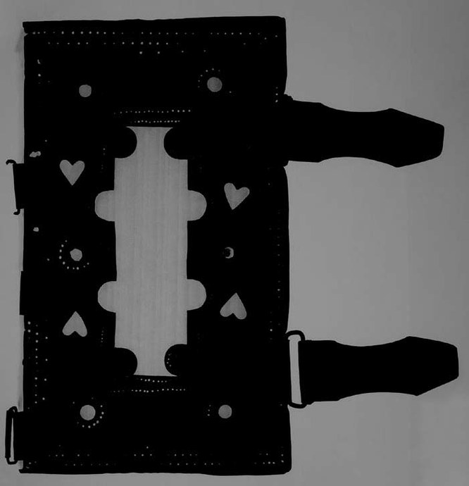
Fig. 3 Separate wooden book cover partly coated with a metal plate. In the area, where metal layer is not placed, there are displayed (invisible by naked eye) year rings of wood itself. The book cover was placed directly on the detector covered with a foil, X-ray tube in distance 49 cm from the detector, voltage on X-ray tube 70 kV, current 300 A, irradiation time for image acquisition 1s. Graphic modification: cut off; retouch by the help of tools patch and pointed retouch brush; gamma correction 1.50; exposition +2.00; contrast 100 (application on the internal structure).
Nebeklíč, 19th century.
Fig. 4 Front board of the book. The bard is coated with bone slices, the sculpture is held in place with little metal nails. The book was placed 36 cm above the detector coated with a foil, X-ray tube in distance 64 cm from the detector, voltage on X-ray tube 110 kV, current 300 A, irradiation time for image acquisition 1 s. Graphic modification: cut off; levels: shift of white; brightness -30; contrast 100.
Albacha Posvátní zvukové, P. J. Herčík, 2nd half of 19th century.
3.2 Book back
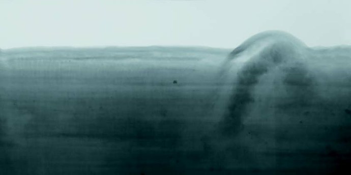
Fig. 5 Book back sewn on raised bands. Ligaments themselves are formed of a simple cord. Seal up with strips of gauze is perceptible between the ligaments. Year rings are also well perceptible on wooden boards. The book placed 36 cm above the detector coated with a foil, X-ray tube in distance 49 cm from the detector, voltage on X-ray tube 90 kV, current 300 A, irradiation time for image acquisition 1 s. Graphic modification: cut off; retouch by the help of tools patch and pointed retouch brush; levels: shift of white; brightness 32; contrast 83.
Animadversiones in regulas et usum critices, R. P. Honorato and S. Maria, 1751.
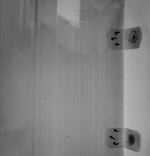
Fig. 6 Book back with false ligaments. Incision for sewing in the backbone is distinctly visible, which is positioned outside placing of strips of false ligaments. The book placed 66 cm above the detector coated a foil, X-ray tube in distance 79 cm from the detector, voltage on X-ray tube 110 kV, current 300 A, irradiation time for image acquisition 1 s. Graphic modification: cut off; brightness 150; contrast 14; levels: shift of white.
Odyssey of Homer, William Cowper, 1855.
3.3 Clips
Fig. 7 Image of vertically placed book. Clips are fixed to the book cover with bent metal wire. The book placed directly on the detector coated with a foil, X-ray tube in distance 49 cm from the detector, voltage on X-ray tube 120 kV, current 300 A, irradiation time for image acquisition 1 s. Graphic modification: cut off; gamma correction 2.00; exposition +1.50.
Pomněnky ve vínek nebeský, Václav Beneš Třebízský, 19th century.
Fig. 8 Strips of clips of book cover. Damage of the wooden board with leather strips from perforated clips is perceptible in the upper part of the image. Migration of iron corrosion products and also fixation of strips by the help of little nails are perceptible in leather. The book placed directly on the detector coated with a foil, X-ray tube in distance 49 cm from the detector, voltage on X-ray tube 75 kV, current 300 A, irradiation time for image acquisition 1 s. Graphic modification: cut off; levels: shift of white; brightness 33; contrast 100; curves: increasing contrast. Animadversiones in regulas et usum critices, R. P. Honorato and S. Maria, 1751.
4. Methods of graphic presentation of results in more complicated cases
In some cases, when displaying units invisible by naked eye, it is difficult to determine their size, especially if there are not displayed any of its components of known sizes in a part of the book shown in the image. This problem can be solved by application of a scale manufactured specifically for purposes of radiography, as it is shown in Fig. 9.
Fig. 9 A hidden element in the front board of the book. The book (together with gauge with smallest division of 2 mm as a scale) placed 66 cm above the detector with coated foil, X-ray tube in distance 79 cm from the detector, voltage on X-ray tube 105 kV, current 300 A, irradiation time for image acquisition 1 s. Graphic modification: cut off; retouch by the help of tools patch and pointed retouch brush; gamma correction 1.50; exposition +1.10; brightness1 20; contrast1 100; contrast2 20.
Za černožlutou oponou (Behind black and yellow curtain), Jaroslav Kunz, 1st half of 20th century.
Conclusions
Non-destructive examination by the help of radiography will help non-destructively detect problematic material and its condition earlier, than serious damage occurs not only in material itself, but of the entire object – a book. Information acquired in this way serves for historical, artistic, scientific understanding of bookbinding. Radiography thus becomes an important tool for obtaining pieces of knowledge on bookbinding and its physical condition without destructive intervention. The method can be generally considered as safe for radiographed materials of bookbinding. Direct displaying follows only dispersion of particles in dependency on chemical composition of the material under examination. Parts of the object, which photons pass through without changes of energy, are shown as light spots in direct displaying (extent of interaction can be judged by comparison with surroundings of the object, where particles have nothing to interact with) – which is mostly just the case of paper and other organic materials, which do not contain heavier elements (other than C, N, O, H). Darker spots represent parts of the object containing heavy elements – classically metals (Fe, Cu …), they present areas, where interaction of primary particles of radiation occurs as well as loss of their original energy. The higher beam voltage, the smaller interaction with the material. A particle, which has kinetic energy high enough, will pass through the material without interacting with it anyhow – without loss of its energy. A particle with lower energy and longer wavelength, typically radiation of light, which the material is capable to absorb, calls up greater response with undesirable manifestations. Radiography thus represents for organic materials smaller stress than investigation in other wavelength of radiation.
In the paper, possibilities of the method are summarized, its parameters, but final evaluation of the method is possible only after research; now detection possibilities are pursued and influence upon radiographed materials. The objective of this paper is to introduce the particular method, not to compare it with other methods or describe them. Comparison of imaging methods will be a part of more extensive work, for example certified methods, as an output of the five-year experimental project. Testing of the effect of x-ray radiation on materials of bookbinding is a part of the partial phase of the five-year experimental project, which is still in progress. At present, possibilities of displaying individual materials are tested, and under what conditions it is possible to obtain relevant information. Examples of particular results acquired by this method are presented in chapter "Typical examples of suitable displaying components of bookbinding" – characteristic elements and defects could not be observable without utilization of this method.
Acknowledgment
This paper was created within the frame of endowment programme of the Ministry of Culture CR, NAKI II No. DG18P02OVV024 in particular, with the title "Use of imaging techniques for the study of hidden information in bookbinding" (2018–2022).
VÁVROVÁ, Petra, NEORALOVÁ, Jitka, HŘEBECKÁ Dana, KOHOUTOVÁ, Kristýna, KULÍČKOVÁ, Anna, MATYSOVÁ, Marie, POPELKOVÁ, Daniela and Tomáš BLECHA.
Non-destructive survey of internal structure of bookbinding by the help of x-ray radiation. Library: Librarian review, 2020, 31(1), …, ISSN 1801- 3252.
References
ALFELD, Matthias, et al. A mobile instrument for in situ scanning macro-XRF investigation of historical paintings. Journal of Analytical Atomic Spectrometry, 2013, 28.5: 760–767.
ALFELD, Matthias, et al. Optimization of mobile scanning macro-XRF systems for the in situ investigation of historical paintings. Journal of Analytical Atomic Spectrometry, 2011, 26.5: 899–909.
CREAGH, D. C. and David A. BRADLEY. Radiation in Art and Archeometry. Elsevier, 2000.
DUIVENVOORDEN, Jorien R., et al. Hidden library: visualizing fragments of medieval manuscripts in early-modern bookbindings with mobile macro-XRF scanner. Heritage Science, 2017, 5.1: 6.
FIALA, P., P. KOŇAS, M. FRIEDL, R. KUBÁSEK and P. ŠMÍRA. X-ray Diagnostics of Wood Invaded by Insect [online]. FEEC VUTBR, 2013 [cit. 16. 10. 2019]. Dostupné z: http://www.measurement.sk/M2013/doc/proceedings/303_Kubasek-2.pdf .
HRADILOVÁ, J., D. HRADIL, O. TRMALOVÁ and J. ŽEMLIČKA, J. Metodika pro vizualizaci vnitřní struktury malířského díla s využitím nových metod na bázi rentgenového záření [online]. Laboratoř ALMA, Akademie výtvarných umění v Praze, Ústav technické a experimentální fyziky ČVUT, 2015 [cit. 16. 10. 2019]. (In Czech: Methods for visualization of internal structure of paintings with utilization of new methods on the basis of x-ray radiation [online]. ALMA laboratory, Academy of beaux arts in Prague, Institute of technical and experimental physics, Czech Technical University, 2015 [ref. 16th Oct., 2019]). Available in: http://invenio.nusl.cz/record/203455/files/nusl-203455_1.pdf .
OSTERLOH, Kurt RS, et al. Fast neutron radiography and tomography of wood as compared to photon based technologies. In: Proceedings of DIR 2007 – International Symposium on Digital Industrial Radiology and Computed Tomography, Lyon, France. 2007. p. 25–27.
PIETIKÄINEN, Markku. Detection of knots in logs using x-ray imaging. VTT, Technical Research Centre of Finland, 1996.
POUYET, E., et al. Revealing the biography of a hidden medieval manuscript using synchrotron and conventional imaging techniques. Analytica chimica acta, 2017, 982: 20–30.
REDO-SANCHEZ, Albert, et al. Terahertz time-gated spectral imaging for content extraction through layered structures. Nature Communications, 2016, 7: 12665.
SCHREINER, M. and H. HOLLE. Documentation of Watermarks in Paper [online]. Institute of Sciences and Technology in Art a Institute of Conservation-Restoration, Academy of Fine Arts, Vienna, 2011 [cit. 16. 10. 2019]. Available in: http://www.restauratorenohnegrenzen.eu/erc/Publications/documents/ERC%20Newsletter_1_2011.pdf
TROJEK, T. and D. TROJKOVA. Several approaches to the investigation of paintings with the use of portable X-ray fluorescence analysis. Radiation Physics and Chemistry, 2015, 116: 321–325.
VAN AKEN, J. An improvement in Grenz radiography of paper to record watermarks, chain and laid lines. Studies in conservation, 2003, 48.2: 103–110.
VAN STAALDUINEN, Mark, et al. Comparing x-ray and backlight imaging for paper structure visualization. EVA-Electronic Imaging & Visual Arts, 2006, 108–113.